Gallery

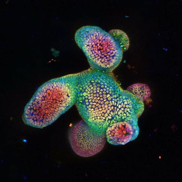
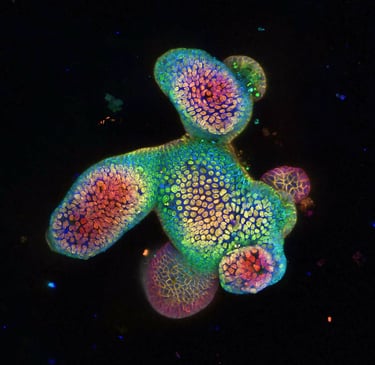
Close-up immunofluorescence microscopy image showing vibrant gut tissue cells stained in green and red against a dark background.
Detailed fluorescent image highlighting immune cell interactions within inflamed intestinal tissue.
Microscopic view of gut lining with bright blue and yellow markers illuminating cellular structures.
High-resolution immunofluorescence image capturing the complex architecture of gut mucosa.
Fluorescent microscopy image revealing protein expression patterns in IBD-affected gut samples.
Vivid immunofluorescence snapshot showing cellular responses in gut tissue under experimental conditions.

Immunofluorescence images from our IBD gut science lab

Contact

We are a team science lab; reach out for collaborations or inquiries @ the e-mail listed at Stanford profiles

Links

Stanford profiles: Michael Schumacher, PhD